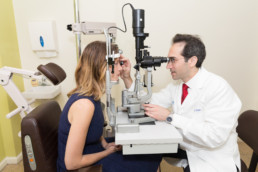
Diagnostic Testing
Visualizing the retina is a fundamental component of the evaluation, and in recent years several new imaging modalities have been integrated into the management of retinal diseases. The physician employs different tests based on the condition that is under investigation.

Ocular Coherence Tomography
With ocular coherence tomography (OCT), your ophthalmologist can see each of the retina’s distinctive layers. This allows your ophthalmologist to map and measure their thickness. These measurements help with diagnosis. They also provide treatment guidance for glaucoma and diseases of the retina. These retinal diseases include age-related macular degeneration (AMD) and diabetic eye disease.
Fundus Photography
Color Fundus Retinal Photography uses a fundus camera to record color images of the condition of the interior surface of the eye, in order to document the presence of disorders and monitor their change over time.
A fundus camera or retinal camera is a specialized low power microscope with an attached camera designed to photograph the interior surface of the eye, including the retina, retinal vasculature, optic disc, macula, and posterior pole (i.e. the fundus).
The retina is imaged to document conditions such as diabetic retinopathy, age related macular degeneration, macular edema and retinal detachment.
Fundus photography is also used to help interpret fluorescein angiography as certain retinal landmarks visible in fundus photography are not visible on a fluorescein angiogram.
Your eyes will be dilated before the procedure. Widening (dilating) a patients pupil increases the angle of observation. This allows the technicians to image a much greater area and have a clearer view of the back of the eye.
Fluorescein Angiography
Fluorescein angiography (FA) is when your ophthalmologist uses a special camera to take pictures of your retina. These pictures help your ophthalmologist get a better look at the blood vessels and other structures in the back of the eye.
Ocular Ultrasound
B-scan ultrasonography is a noninvasive and commonly used diagnostic tool in the assessment of a variety of ocular and orbital diseases. With proper use, the technique affords vast information unobtainable through clinical exam alone. The echographic exam of the human eye and orbit is described, and echographic characteristics of various ocular pathologies are illustrated.
